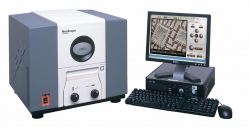
Innovative Implementation: Scanning Electron Microscope (SEM)
In February 2012, the Center for Renewable and Alternative Fuel Technologies (CRAFT) team obtained a lab instrument that will provide new and exciting research capabilities for its Cellulosic-Derived Biofuels Initiative. CRAFT’s new Scanning Electron Microscope (SEM) goes far above the capabilities of the run-of-the-mill optical microscopes which tend to have a magnification power of around 1,000 times. CRAFT’s SEM has capabilities of magnification up to 40,000 times in 3D. SEMs also have tremendous depth of field compared to conventional microscopes, providing an almost 3D image for researchers to analyze, as compared to the flatter image an optical microscope produces. Additionally, these advanced microscopes can look past the surface of an object, telling researchers information about its composition.
So, how does an SEM work?
Scanning electron microscopes utilize electrons in place of light beams to achieve both higher magnification and greater clarity and depth of field over traditional microscopes. Preparing a sample of examination by an SEM is a complicated process. First, the sample must be dehydrated because it will be placed in a vacuum chamber. If it is a non-metal, it must then be covered with a thin conductive covering to prevent "charging" by the electrons, usually gold foil. This process is called "sputter coating."
A beam of electrons is produced from the "electron gun" located at the top of the SEM. These electrons are usually produced by the heating of a metallic filament like tungsten. The electron beam from the "gun" follows a path down through the microscope past electromagnetic lenses which focus the beam towards the sample. When the beam hits the sample, other electrons are dispersed or ejected from the sample. These electrons are called back scattered or secondary electrons. Specialized detectors placed in the SEM collect the ejected electrons and convert them to a signal sent to a viewing screen. The screen assembles the signal into an image. The images produced are that of the quality found in scientific journals and educational text books.
Read more: How Does an SEM Microscope Work?
EKU Faculty and Student Benefits of the SEM
CRAFT’s SEM is the first of its kind on EKU’s campus. According to CRAFT Program Director, Bruce Pratt, “Faculty can expand their research by designing experiments to take advantages of the capabilities of the SEM. Experiments can be designed to examine physical characteristics in much more detail than would be possible with a light microscope. It also gives a 3D image of the material being examined.
Several students have been trained on the SEM and will have the opportunity to use a state of the art piece of analytical equipment. CRAFT Research Technician, Gary Selby explains: “One of my students (Nan Campbell) acquired a skill that could be valuable as she begins her career. Dr. Pratt has made it clear that he wants the SEM to be used and that it won't be limited to CRAFT research. This means that the entire university could benefit from the SEM if they choose.”
The Use of the SEM at CRAFT
The main focus of this SEM in CRAFT research will focus on looking at physical changes in different types of biomass during the chemical pretreatment process of biomass for biofuels production. By looking at how the different pre-treatment processes physically disrupt the biomass, CRAFT researchers can better optimize the process of getting sugars from our biomass for fermentation by micro-organisms. The SEM allows for researchers to rapidly assess the level of degradation of biomass after pretreatment and saccharification.
(Above, you can see a picture of switchgrass taken by the SEM pretreated with NaOH at 4,000X magnification.)
The SEM is currently being used on a study to compare physical degradation of biomass after pretreatment, to sugars released after saccharification. “We also plan to look at algae at different stages of growth in the future,” says Pratt.
Published on April 16, 2012

.gif)